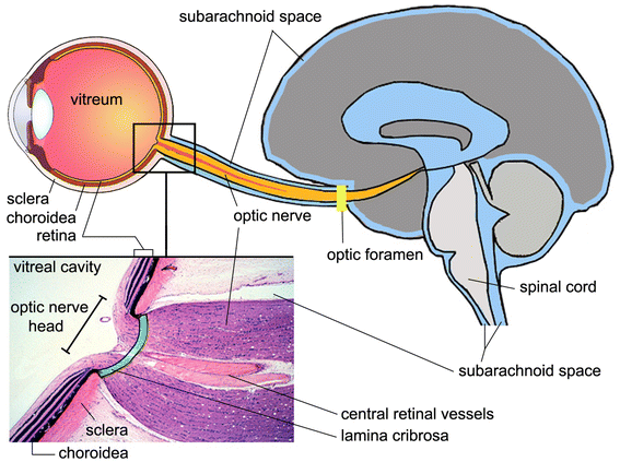
Figure 1
From: A new glaucoma hypothesis: a role of glymphatic system dysfunction

Section of eye, optic nerve head and brain, illustrating the subarachnoid space. The lamina cribrosa (green) is a sieve-like structure in the posterior part of the sclera and allows the passage of the retinal ganglion cell axons and the central retinal vessels. The optic nerve is surrounded by cerebrospinal fluid (blue) in the subarachnoid space. The enlarged histologic view is modified and reproduced with permission, Boston University Histology Learning System (Deborah W. Vaughan, PhD).
